.jpg)
Surface Modifications of Dental Implants for Enhanced Antimicrobial Properties: A Literature Review
Yuval Reiser 1  , Marko Vuletic2 and Dragana Gabric2*
1School of Dental Medicine, University of Zagreb, , Zagreb, Croatia .
2Department of Oral Surgery, School of Dental Medicine, University of Zagreb; Clinical Hospital Center Zagreb, University Dental Clinic, Zagreb Croatia .
http://dx.doi.org/10.12944/EDJ.07.0102.04
Dental implants are widely used in modern dentistry for tooth replacement, but bacterial infections such as peri-implant mucositis and peri-implantitis remain significant causes of implant failure. This literature review evaluates the effectiveness of surface modifications and antimicrobial coatings in reducing bacterial colonization and infection on dental implants. A comprehensive search of PubMed and Google Scholar identified 70 relevant studies, of which 56 met the inclusion criteria. The review highlights the use of antimicrobial agents such as silver, zinc, copper, fluorine, and chlorhexidine, as well as surface modifications like dendrimers, titanium dioxide photocatalysts, and ultraviolet treatment. These strategies enhance antimicrobial properties by generating reactive oxygen species, creating super-hydrophilic surfaces, and altering surface characteristics such as roughness, hydrophobicity, charge, and crystalline phase. The findings suggest that these approaches significantly reduce bacterial adhesion and biofilm formation, thereby improving implant success rates. However, further research is needed to assess the long-term clinical performance and stability of these modifications. This review highlights the crucial role of combining advanced surface modification techniques with antimicrobial approaches to improve the performance and safety of dental implants.
Copy the following to cite this article:
Reiser Y, Vuletic M, Gabric D. Surface Modifications of Dental Implants for Enhanced Antimicrobial Properties: A Literature Review. Enviro Dental Journal 2025; 7(1).
DOI:http://dx.doi.org/10.12944/EDJ.07.0102.04Copy the following to cite this URL:
Reiser Y, Vuletic M, Gabric D. Surface Modifications of Dental Implants for Enhanced Antimicrobial Properties: A Literature Review. Enviro Dental Journal 2025; 7(1).Avialable here: https://bit.ly/3Mn1WDE
Download article (pdf) Citation Manager

Introduction
In dental implantology, bacterial diseases of oral cavities, such as peri-implant mucositis and peri-implantitis, can result in loss of implants. Dental implants offer sub- and supragingivally located surfaces that are available to bacterial colonization. The microbiological findings show clearly that infection represents a possible etiological factor for late changes in implants 1-3. Peri-implant mucositis, characterized by reversible inflammation of the soft tissues around the implant, can progress to peri-implantitis if left untreated. Peri-implantitis involves not only soft tissue inflammation but also bone resorption, leading to implant instability and potential loss 3. Beyond local effects, systemic consequences of bacterial translocation from the oral cavity are increasingly recognized. Bacteria that enter the bloodstream during dental procedures, including dental implant therapy, can potentially lead to severe systemic infections such as infective endocarditis 4.
Bacteria residing in biofilm such as anaerobic gram-negative species including Porphyromonas gingivalis, Prevotella intermedia, and Tannerella forsythia, along with facultative anaerobes like Staphylococcus aureus, are difficult to eliminate using systemic antibiotics. To address this challenge, various anti-adhesive and antimicrobial approaches have been developed to hinder the development of mature biofilms on biomaterial surfaces by minimizing or preventing the initial stages of bacterial attachment and colonization 5. It includes surface coatings with dendrimers or polymeric brushes, such as poly(ethylene glycol) layers 6,7, surface modifications giving positive charges or turning the surface into a zwitterion layer 8,9, titanium implant
Surface antimicrobial coatings encompass various types, such as ionic antibacterial layers, antibiotic or organic-based antimicrobial coatings, and substrates embedded with antibacterial compounds. Additionally, surface treatments utilizing silver nanoparticles or photocatalytic materials have also been used for antimicrobial purposes 5.
Titanium implant surface modifications can provide antimicrobial materials and offer a means to superimpose bacterial adhesion or biosynthesis by changing the surface wettability, texture, crystal morphology, surface potential and other characteristics 10.
The aim of this literature review is to explain the benefits of dental implant surface modifications acting on opportunistic microorganisms and pathogenic bacteria that can lead to an infection and thus to dental implant failure.
Materials and Methods
Scope: This review explores recent advancements in surface modification techniques for dental implants that enhance antimicrobial properties, including ionic antibacterial coating, antibiotics and organic antimicrobial coating and surface modifications, aiming to reduce bacterial adhesion and improve implant success.
Protocol development
Three authors (Y.R, M.V and D.G) used PICO system to formulate, present and answer the question: How
do the surface modifications of dental implants affect the antimicrobial properties in implant patients?
Participants (P): Patients receiving dental implants.
Intervention or Exposure and Comparison Group (IC): Surface modifications\coating of dental providing antimicrobial properties. Comparator: implants that did not go through surface modifications or coating prior implantation.
Outcome (O): Reduction in bacterial adhesion, colonization or biofilm formation.
Eligibility criteria
Inclusion criteria
Dental implantology
Publications in English
Antibacterial or anti-adhesive properties of the dental implant surface
Dental implant surface modification or coating
Exclusion criteria
No surface modification or coating
No relation to implantology
Search strategy
Two electronic databases were searched and information gathered from November 2 until December 17, 2024, as shown in table 1. In order to identify articles in the topic of interest, several related terms were used: “Dental implant surface modifications”, “Antimicrobial effect of dental implant surface coating” and “antimicrobial effect of dental implant surface modifications”.
Publications from 1974 up to 2024 in the interest topic were gathered, duplicates were removed manually, following which the titles and abstracts of the articles were screened independently based on the predefined inclusion and exclusion criteria.
Table 1: Electronic databases and search terms.
Electronic database | Search term |
PubMed | “Dental implant surface modifications”, “Antimicrobial effect of dental implant surface coating”, “antimicrobial effect of dental implant surface modifications”. |
Google Scholar | “Dental implant surface modifications”, “Antimicrobial effect of dental implant surface coating” and “antimicrobial effect of dental implant surface modifications”. |
Data extraction
Relevant data were individually extracted from the selected articles by three reviewers (Y.R, M.V and D.G) using separate customized charts. Information such as authors' names, year of publication, and other data relevant to the topic were categorized and assessed within the charts. A condensed overview of the included studies is presented in Table 2, while the complete dataset, detailing all studies and their characteristics, is provided in the Supplementary Material (Table S1).
Table 2: Included study characteristics.
Type | Number of studies | Key points\ Main findings |
Microbiology | 7 | Investigated microbial causes of implant infections such as peri-implant mucositis and peri-implantitis. Identified contamination sources including implant surfaces, surgical staff, and patient flora. Highlighted systemic risks (e.g., bacteremia, infective endocarditis). |
Ionic Antibacterial coating | 16 | Explored metallic coatings with Ag, Zn, Cu, and F ions. Silver and zinc coatings showed strong antimicrobial activity through ion release, reactive oxygen species, and photocatalysis while maintaining biocompatibility. Copper exhibited antimicrobial and anti-inflammatory effects. Fluoride coatings (e.g., TiF4) inhibited Streptococcus mutans and Bacteroides gingivalis. |
Antibiotics and organic antibiotics coating | 15 | Studied coatings using antibiotics (amoxicillin, vancomycin, gentamicin, etc.), antimicrobial peptides (AMPs), dextran, polyethylene glycol (PEG), and chlorhexidine. AMPs provided broad-spectrum antibacterial activity with low resistance risk. PEG/dextran coatings reduced protein, platelet, and bacterial adhesion. Chlorhexidine coatings achieved sustained antimicrobial release on titanium surfaces. |
Surface modifications | 25 | Examined physical and chemical surface treatments such as TiO2 coatings, UV photo-functionalization, nano structuring, charge alteration, and hydrophilicity tuning. Findings showed that super-hydrophilic, negatively charged, and nano-textured Ti surfaces decrease bacterial adhesion and biofilm formation. The crystalline phase of TiO2 (anatase, rutile, brookite) influenced photocatalytic antibacterial effects. |
Results
A Flowchart was constructed using PRISMA flow diagram showing the identification, screening and selection of published materials for this review paper (see Appendix A).A total of 70 articles were found from electronic database search (PubMed and Google Scholar). Overall, 3 duplicates were removed, and 11 articles were excluded based on the inclusion and exclusion criteria. 56 publications were selected for abstract and full text analysis.
Ionic antibacterial coating
A total of 16 publications were selected for this review. Among them, five studies focused on silver coatings for implants, six on zinc coatings, three on copper coatings, and two on fluorine coatings. Additionally, four publications did not meet the inclusion criteria and were excluded. The implant is packaged in a sterile, air-tight container, with infections being rare due to strict medical device industry controls 10. Infections can arise from contaminated implant surfaces, surgical staff, the patient’s skin or mucosa, remote infections, contaminated disinfectants, or contact with others post-procedure 11,12. Silver (Ag) is a powerful antimicrobial, acting through Ag+ ions or "contact killing," which disrupts bacterial cell walls and membranes, leading to cell death 13,14. Techniques for silver antimicrobial coatings include plasma spraying and electrochemical deposition 9. Radtke et al. 15 developed a silver-doped titanium oxide nanotube coating, achieving 97.62% bacterial inhibition via hydroxyl radicals. Silver nanoparticles (AgNPs) enhance antimicrobial effects due to their large surface area and stable oxidative stress reactions 16. Zinc offers antimicrobial properties through ion, contact, and photocatalytic mechanisms, disrupting bacterial adhesion and membranes 17-21. Wang et al. 22 developed a zinc-doped ZrO2/TiO2 coating with strong antimicrobial effects against Staphylococcus aureus and good biocompatibility. Copper has anti-inflammatory and antimicrobial properties and is safe at proper levels 23. Jannesari et al. 24 showed copper ions inhibit bacterial respiration and DNA. Fluorine, zinc, and copper can be added to titanium or hydroxyapatite coatings, with antimicrobial effects from gradual ion release 25. Titanium tetrafluoride (TiF4) inhibits Streptococcus mutans and Bacteroides gingivalis on teeth 26, and fluoride ions on titanium surfaces effectively combat peri-implantitis bacteria 27.
Antibiotics and organic antimicrobial coating
In the context of antimicrobial coatings, 15 publications were included in the review. Of these, eight studies examined surface treatments using conventional antibiotics and antimicrobial peptides, five focused on dextran and polyethylene glycol (PEG), and two investigated chlorhexidine. One publication did not meet the inclusion criteria and was excluded. Conventional antibiotics are used in controlled release devices but can cause resistance due to an initial burst and prolonged sub- minimal inhibitory concentration (MIC) release 25. To address this, antimicrobial peptides (AMPs) offer broad-spectrum activity and reduce resistance risks by binding to bacterial membranes and targeting intracellular components 28-32. Various coating methods, including adsorption and electrospinning, help prevent bacterial colonization on implants 33,34. Dextran and polyethylene glycol (PEG) coatings inhibit adhesion of proteins, platelets, and several types of bacteria 35-38. Ion-implanted surfaces showed 55-80% fewer bacteria than pure titanium 39. Chlorhexidine (CHX) is effective against gram-positive and gram-negative bacteria in oral care 40. Wood et al. 41 developed a CHX nanoparticle coating with sustained antimicrobial effects against Streptococcus gordonii within 8 hours.
Surface modifications
Surface modifications emerged as the most extensively studied topic, with 25 publications included in this review. Among these, three studies explored surface treatments with titanium dioxide, three investigated ultraviolet (UV) treatment, four focused on surface roughness, six examined surface charge, three analyzed surface hydrophilicity, and six assessed the impact of surface crystalline phase on antimicrobial properties. Six publications were excluded for not meeting the inclusion criteria. Pure titanium forms a biocompatible and stable titanium dioxide (TiO2) layer in air, supporting osseointegration and offering antimicrobial properties under certain conditions 42. TiO2 nanotubes reduce bacterial adhesion after ultraviolet (UV) exposure, while TiO2 nanoparticles damage bacterial membranes via reactive oxygen species (ROS) 43. TiO2 can also serve as a carrier for sustained drug release 9, and UV treatment decreases bacterial adhesion on implants 44. Suketa et al. 45 showed that an anatase TiO2 layer on titanium reduced Actinobacillus actinomycetemcomitans and Fusobacterium nucleatum viability to under 1% with ultraviolet A (UVA) exposure. UV-induced TiO2 photo-functionalization creates a super-hydrophilic surface that oxidizes organic impurities for antimicrobial effects 46. Protein adsorption on implant surfaces is influenced by hydrophobicity, roughness, and chemical composition, affecting bacterial adhesion 47-50. Bacteria often adhere to protein-coated surfaces, leading to biofilm formation through gene expression changes 51-53. Combining biofilm-disrupting agents with antibiotics may improve outcomes, though this is rarely practiced due to chemical aggressiveness 54. Surface smoothness reduces microbial colonization, but nano roughness can enhance protein adsorption and reduce bacterial adhesion 5,54. Rough surfaces increases plaque accumulation in dentistry 55. Techniques like ion implantation, laser cladding, and anodizing create nano-scale structures 56,57. Surface charge and hydrophilicity affect bacterial adhesion; negatively charged and hydrophilic surfaces generally reduce bacterial attachment 51,52,57,58. Bacterial adhesion depends on electrostatic interactions and substrate-bacteria physicochemical properties 59. Gao et al. 60 linked antibacterial effects to electrostatic disturbances causing cell death. Hydrophobic interactions drive adhesion of pathogens like Streptococcus aureus and Staphylococcus epidermidis 61,62. TiO2’s crystalline forms like anatase, rutile, and brookite, all contribute to antibacterial properties through photocatalytic activity (PCA) 54,64-66.
Across all modalities, ion-based coating provided the strongest broad-spectrum antimicrobial effect. Peptide-based coating showed excellent biofilm prevention with a low risk for antibiotic resistance. Surface roughness and charge modification improved early bacterial suppression but required combination with other modalities for stronger antimicrobial performance.
Discussion
Dental implant infections are a rare but serious complication that can arise from various sources, including contaminated implant surfaces, surgical staff, or the patient’s own skin. Silver is highly effective in combating microbial contamination through ion release or direct contact killing, and silver-based coatings, including silver nanoparticles, have been developed for enhanced antimicrobial activity. Similarly, other metals like zinc and copper exhibit strong antimicrobial properties through various mechanisms, with some coatings also maintaining biocompatibility. Antimicrobial peptides offer a promising alternative to antibiotics, as they exhibit broad-spectrum activity and are less prone to inducing bacterial resistance. Titanium dioxide, commonly used in implants for its biocompatibility and photocatalytic properties, can also be utilized to reduce bacterial adhesion, especially under ultraviolet light. Furthermore, surface modifications such as nanopatterning and doping with antimicrobial agents can significantly reduce bacterial colonization and biofilm formation, thereby improving the longevity and safety of dental implants.
Ionic antibacterial coating
Silver (Ag) stands out as the most potent metal against microbes, exerting its antibacterial activity either through the release of Ag+ ions or by directly destroying microorganisms upon contact 13,14. Various techniques have been developed to create silver-containing antimicrobial coatings, including plasma spraying, electrochemical deposition, sol-gel processes, thermal spraying, co-sputtering, and plasma immersion ion implantation 9. Radtke A et al. 15 developed a silver-doped titanium oxide nanotube array coating by immersing the substrate in a silver nitrate solution. This modification effectively suppressed biofilm formation Silver nanoparticles (AgNPs) have also been recognized as a promising alternative for silver-based antimicrobial coatings. AgNPs provide long-lasting and stable antimicrobial activity through oxidative stress mechanisms and the generation of reactive oxygen species16. Similar to silver, zinc exerts its antimicrobial properties through the three mechanisms earlier described 17-19. Wang et al. 22 developed a zinc-doped ZrO2/TiO2 coating on a Titanium/Aluminum/Vanadium alloy (Ti6Al4V) surface using a combination of magnetron sputtering and microarc oxidation. This coating demonstrated exceptional antimicrobial effectiveness against Staphylococcus aureus while maintaining biocompatibility and corrosion resistance. Copper is an inexpensive and readily available metal that has been utilized since ancient times, both in its pure form and as metal compounds, for its anti-inflammatory, antimicrobial, and anti-proliferative properties 23. At appropriate levels, copper is non-toxic to humans. Research by Jannesari et al. 24 indicated that copper ions and their superoxide derivatives can disrupt bacterial respiratory processes and cause degradation of bacterial DNA, thus providing significant antimicrobial effects. Elements such as fluorine (F), zinc (Zn), and copper (Cu) can be integrated into titanium or hydroxyapatite coatings through anodic oxidation of their respective ions. The bactericidal action of these ions appears to rely on their gradual release into the surrounding environment, with one proposed mechanism for bacteriostasis being the hydroxylation into highly reactive components 25. In vitro study by Skartveit et al. 26 demonstrated that titanium tetrafluoride (TiF4) effectively inhibits the proliferation of Streptococcus mutans and Bacteroides gingivalis on tooth surfaces. Meanwhile, Yoshinari et al. 27 successfully introduced fluoride ions to the surface of pure titanium using ion accelerators, achieving effective inhibition of pathogenic bacteria associated with peri-implantitis.
Antibiotics and organic antimicrobial coating
Conventional drug studies have utilized antibiotics such as amoxicillin, vancomycin, gentamicin, tetracycline, minocycline, and cephalothin, incorporated into controlled-release systems. A major issue with these antibiotics is the initial burst release, which is often followed by a sustained release at levels below the MIC, potentially promoting the development of bacterial resistance over time 25. Due to their tendency to promote drug resistance, the use of antibiotics is limited, whereas the development of AMPs offers a new strategy to address this challenge 28. AMPs are short amphiphilic peptides with broad-spectrum antimicrobial activity. Their positive charge allows them to interact with negatively charged bacterial membranes via electrostatic attraction, ultimately resulting in bacterial cell death 29,30. Research has shown that AMPs can penetrate the bacterial cytoplasm to target intracellular components such as DNA, RNA, and proteins, thereby killing bacteria from within and affecting gene expression 31,32. Due to their unique antibacterial mechanisms, bacteria find it challenging to develop resistance. Various methods have been employed to coat antimicrobial peptides onto implant surfaces to prevent bacterial colonization, including adsorption, binding, electrospinning, and chemical attachment 33,34. Dextran and polyethylene glycol (PEG) surfaces have proven effective at inhibiting protein adhesion, platelet adhesion, bacterial adhesion, and biofilm formation by oral pathogens such as Staphylococcus aureus, Streptococcus sanguinis, Lactobacillus salivarius, Streptococcus mutans, and Streptococcus gordonii 35-38. Bacterial counts on ion-implanted surfaces decreased significantly compared to pure titanium 39. Chlorhexidine (CHX) is a widely utilized surface disinfectant known for its effectiveness against both gram-positive and gram-negative bacteria. Consequently, it is frequently employed for local cleaning and disinfection during oral surgical procedures and oral care 40. Wood et al. 41 developed a chlorhexamethy hexameta-phosphate nanoparticle coating on pure titanium, with an average particle diameter of 49 nm. This coating demonstrated sustained release of CHX and exhibited antimicrobial activity against the oral primary colonizing bacterium Streptococcus gordonii within 8 hours.
Surface modifications
Pure titanium rapidly develops a thin layer of titanium dioxide (TiO2) when exposed to air, which exhibits excellent biocompatibility and chemical stability. Additionally, TiO2 possesses antimicrobial properties under certain conditions 42. For instance, TiO2 nanotubes can significantly inhibit bacterial adhesion post-ultraviolet radiation exposure, while TiO2 nanoparticles can damage bacterial membranes by generating ROS 43. Furthermore, TiO2 can act as a carrier for antimicrobial drugs, allowing for their sustained release from the metal 9. Bacterial adhesion and polymerization on implant surfaces significantly decreased after 8 hours of UV treatment 44. In research conducted by Suketa et al. 45, a thin layer of photocatalytic anatase TiO2 was applied to the surface of pure titanium via plasma source ion implantation followed by annealing. This layer demonstrated a robust photocatalytic reaction under UVA illumination, suppressing the viability of both Actinobacillus actinomycetemcomitans and Fusobacterium nucleatum to less than 1% under UVA exposure within 120 minutes. The UV light-induced photo-functionalization of TiO2 eliminates hydrocarbon contamination and results in a super-hydrophilic surface that oxidizes adsorbed organic impurities. This secondary oxidation, initiated by ROS, is believed to be essential for achieving antimicrobial activity 46. Protein adsorption is influenced by several factors, including surface hydrophobicity, roughness, porosity, chemical composition, as well as the composition and concentration of the protein solution, salt concentrations, and pH 47-50. The layer of adsorbed proteins is crucial for bacterial adhesion. In reality, bacteria rarely encounter a "clean" surface on an implant. Free-swimming bacteria, in the so-called planktonic state, tend to adhere to these surface-adsorbed proteins 51,52. The adhered bacteria can proliferate and attract other bacteria from the surrounding environment. Once a sufficient population of bacteria forms a colony on the surface, they will modify their gene expression patterns. Genes responsible for producing extracellular polymeric substances, vital for biofilm formation, will be activated and expressed 52,53. The combined use of biofilm-disrupting agents alongside antibiotics appears to be a more effective strategy; however, this approach is not frequently implemented in practice due to the aggressive nature of the chemicals involved 52. It is evident that the topological and chemical properties of a dental implant surface significantly influence the adhesion rate of microorganisms. A completely smooth surface is less prone to microbial colonization compared to a rough surface, which provides a larger area for adhesion and greater adhesive forces generated by microorganisms per unit area 5. Although, Puckett et al. 54 noted that surfaces with increased nano roughness exhibit higher surface energy, leading to enhanced protein adsorption and, consequently, reduced bacterial adhesion. However, specific threshold values regarding surface roughness that may affect bacterial adhesion remain undefined. In the field of dentistry, a rise in plaque accumulation has been observed with roughness measurements exceeding 0.2 um 55. Various methods are utilized to create nano-scale structures on implant surfaces. Zhang et al. 56 successfully produced a silver-zinc oxide-hydroxyapatite (Ag-ZnO-HA) nanocomposite on Ti6Al4V using laser cladding, which demonstrated excellent antibacterial properties against Escherichia coli and Streptococcus aureus in vitro, along with favorable osteogenesis properties in vivo. Most microorganisms possess a charged outer membrane and contain hydrophobic regions, which may contribute to their adhesion to the hydrophobic surfaces of medical implants. Nonetheless, the adhesion of microorganisms typically relies on the formation of a protein layer on the surface, potentially revealing high-affinity adhesion sites 51,52. Negatively charged surfaces tend to be less prone to bacterial adhesion than positively charged surfaces, as most bacteria have negatively charged cell walls 57,58. The effect of electrical repulsion on bacterial adhesion becomes more pronounced with increased surface hydrophilicity. In terms of surface free energy, the degree of bacterial adhesion can either rise or fall with increasing surface energy of substrates, depending on the physicochemical characteristics of both the substrate and the bacterial strains involved, as well as the aqueous solution utilized 59. Gao et al. 60 found that the antibacterial effect observed on the studied surface resulted from electrostatic disturbances that triggered autolytic and/or cellular death mechanisms. Surface hydrophilicity contributes to the interactions between biomaterials and bacteria, but these systems are complex and shaped by various factors, including the implant location and the specific bacterial species and strains involved. Theoretically, hydrophobic bacterial strains are more likely to adhere to biomaterials with hydrophobic characteristics, while hydrophilic species are inclined to attach to hydrophilic surfaces 61. Hydrophobic interactions are common and play a role in the mechanisms of various microbial adhesion factors, including hydrophobic components of cell membranes and adhesins found on fimbriae or pili 62. Research has confirmed that the adhesion of human pathogens such as Streptococcus aureus and Staphylococcus epidermidis correlates with increased hydrophobicity of the biomaterial surface, establishing hydrophobicity as a primary driver of bacterial adhesion 63. Concerning the crystalline phases of the surface, titanium dioxide can exist in three forms. The crystalline structure is closely linked to its ability to inhibit bacterial adherence 54,64,65. All three forms exhibit photocatalytic activity, which contribute to their antibacterial properties 61,64,66.
All included studies were published in English, which may introduce language bias and limit of the findings. The review primarily focused on in vitro and some in vivo studies, which may not fully reflect clinical outcomes in human subjects. Furthermore, variations in study design, methodologies, and outcome measures among the reviewed studies could impact the consistency and comparability of the results.
Conclusions
This review highlights the critical role of surface modifications and antimicrobial coatings in reducing infections in dental implants. Materials like silver, zinc, copper, and fluorine exhibit strong antimicrobial properties, preventing bacterial growth through ion release, oxidative stress, and photocatalytic activity. Additionally, surface characteristics such as roughness, charge, and crystalline phase significantly influence bacterial adhesion, underscoring the need for optimized implant surface designs. Ultraviolet treatment and titanium dioxide coatings further enhance antimicrobial effects, with titanium dioxide generating reactive oxygen species to disrupt bacterial membranes. Other strategies, including chlorhexidine coatings and controlled-release antibiotic devices, offer infection control but face challenges like bacterial resistance. Meanwhile, alternatives such as dextran and polyethylene glycol coatings effectively reduce biofilm formation and bacterial adhesion, presenting promising solutions for implant-related infections. Despite these advancements, further research is essential to evaluate the long-term stability and performance of these coatings, particularly under different clinical conditions.
Acknowledgement
The authors thank the School of Dental Medicine, University of Zagreb, for institutional support and Dr. Vadim Reiser for his valuable guidance during the preparation of this review.
Funding Sources
The research was funded as part of the project 'Evaluation of Innovative and Standard Protocols in Endodontic and Surgical Therapy', no. SFZG-05- 2025, INOENDO, co-financed from the funds of the National Recovery and Resilience Plan (NRRP).
Conflict of Interest
The authors do not have any conflict of interest.
Authors’ Contribution
Y.R. (Yuval Reiser): Conceptualization, Methodology, Investigation, Writing – Original Draft, Writing-Review and Editing.
M.V. (Marko Vuletic): Software, Validation, Data Curation, Writing – Original Draft, Writing-Review and Editing.
D.G. (Dragana Gabric): Conceptualization, Visualization, Supervision, Validation, Project Administration, Writing-Review and Editing.
References
- Pye AD, Lockhart DE, Dawson MP, Murray CA, Smith AJ. A review of dental implants and infection. J Hosp Infect. 2009;72(2):104–110. doi:10.1016/j.jhin.2009.02.010
CrossRef - Norowski PA Jr, Bumgardner JD. Biomaterial and antibiotic strategies for peri-implantitis: a review. J Biomed Mater Res B Appl Biomater. 2009;88(2):530–543. doi:10.1002/jbm.b.31152
CrossRef - Esposito M, Hirsch JM, Lekholm U, Thomsen P. Biological factors contributing to failures of osseointegrated oral implants. (II). Etiopathogenesis. Eur J Oral Sci. 1998;106(3):721–764. doi:10.1046/j.0909-8836.1998.t01-6-.x
CrossRef - Kerrigan SW, Cox D. Platelet-bacterial interactions. Cell Mol Life Sci. 2009;67(4):513–523. doi:10.1007/s00018-009-0207-z
CrossRef - Knetsch MLW, Koole LH. New strategies in the development of antimicrobial coatings: the example of increasing usage of silver and silver nanoparticles. Polymers (Basel). 2011;3(1):340–366. doi:10.3390/polym3010340
CrossRef - Eichler M, Katzur V, Scheideler L, et al. The impact of dendrimer-grafted modifications to model silicon surfaces on protein adsorption and bacterial adhesion. Biomaterials. 2011;32(38):9168–9179. doi:10.1016/j.biomaterials.2011.08.063
CrossRef - Müller R, Eidt A, Hiller KA, et al. Influences of protein films on antibacterial or bacteria-repellent surface coatings in a model system using silicon wafers. Biomaterials. 2009;30(29):4921–4929. doi:10.1016/j.biomaterials.2009.05.079
CrossRef - Roosjen A, Norde W, van der Mei HC, Busscher HJ. The use of positively charged or low surface free energy coatings versus polymer brushes in controlling biofilm formation. In: Grundke K, Stamm M, Adler HJ, eds. Characterization of Polymer Surfaces and Thin Films. Springer; 2009:132. doi:10.1007/2882_026
CrossRef - Zheng TX, Li W, Gu YY, Zhao D, Qi MC. Classification and research progress of implant surface antimicrobial techniques. J Dent Sci. 2022;17(1):1–7. doi:10.1016/j.jds.2021.08.019
CrossRef - Young WT. How to respond to changes in the regulation of the ethylene-oxide sterilization process. Med Device Technol. 2004;17(3):12–15. PMID:16736657
- Rosenthal VD, Yin R, Nercelles P, et al. International Nosocomial Infection Control Consortium (INICC) report of healthcare-associated infections, data summary of 45 countries for 2015 to 2020, adult and pediatric units, device-associated module. Am J Infect Control. 2024;52(9):1002–1011. doi:10.1016/j.ajic.2023.12.019
CrossRef - Bearman GM, Munro C, Sessler CN, Wenzel RP. Infection control and the prevention of nosocomial infections in the intensive care unit. Semin Respir Crit Care Med. 2006;27(3):310–324. doi:10.1055/s-2006-945534
CrossRef - Wei X, Li Q, Wu C, Sun T, Li X. Preparation, characterization, and antibacterial mechanism of the chitosan coatings modified by Ag/ZnO microspheres. J Sci Food Agric. 2020;100(13):5527–5538. doi:10.1002/jsfa.10605
CrossRef - Bui VD, Mwangi JW, Schubert A. Powder mixed electrical discharge machining for antibacterial coating on titanium implant surfaces. J Manuf Process. 2019;44:261–270. doi:10.1016/j.jmapro.2019.05.032
CrossRef - Radtke A, Topolski A, Jedrzejewski T, et al. The bioactivity and photocatalytic properties of titania nanotube coatings produced with the use of the low-potential anodization of Ti6Al4V alloy surface. Nanomaterials (Basel). 2017;7(8):197. doi:10.3390/nano7080197
CrossRef - Yin IX, Zhang J, Zhao IS, Mei ML, Li Q, Chu CH. The antibacterial mechanism of silver nanoparticles and its application in dentistry. Int J Nanomedicine. 2020;15:2555–2562. doi:10.2147/IJN.S246764
CrossRef - de Lima CO, de Oliveira ALM, Silva Filho EC, et al. Zn-doped mesoporous hydroxyapatites and their antimicrobial properties. Colloids Surf B Biointerfaces. 2020;198:111471. doi:10.1016/j.colsurfb.2020.111471
CrossRef - Maimaiti B, Zhang N, Yan L, et al. Stable ZnO-doped hydroxyapatite nanocoating for anti-infection and osteogenic properties on titanium. Colloids Surf B Biointerfaces. 2019;186:110731. doi:10.1016/j.colsurfb.2019.110731
CrossRef - Padmavathy N, Vijayaraghavan R. Enhanced bioactivity of ZnO nanoparticles—an antimicrobial study. Sci Technol Adv Mater. 2008;9(3):035004. doi:10.1088/1468-6996/9/3/035004
CrossRef - Memarzadeh K, Sharili AS, Huang J, Rawlinson SC, Allaker RP. Nano-particulate zinc oxide as a coating material for orthopedic and dental implants. J Biomed Mater Res A. 2015;103(3):981–989. doi:10.1002/jbm.a.35241
CrossRef - Liu Y, He L, Mustapha A, Li H, Hu ZQ, Lin M. Antibacterial activities of zinc oxide nanoparticles against Escherichia coli O157:H7. J Appl Microbiol. 2009;107(4):1193–1201. doi:10.1111/j.1365-2672.2009.04303.x
CrossRef - Wang R, He X, Gao Y, Zhang X, Yao X, Tang B. Antimicrobial property, cytocompatibility, and corrosion resistance of Zn-doped ZrO2/TiO2 coatings on Ti6Al4V implants. Mater Sci Eng C Mater Biol Appl. 2017;75:7–15. doi:10.1016/j.msec.2017.02.036
CrossRef - Li Y, Liu L, Wan P, et al. Biodegradable Mg-Cu alloy implants with antibacterial activity for the treatment of osteomyelitis: in vitro and in vivo evaluations. Biomaterials. 2016;106:250–263. doi:10.1016/j.biomaterials.2016.08.031
CrossRef - Jannesari M, Akhavan O, Madaah Hosseini HR, Bakhshi B. Graphene/CuO2 nanoshuttles with controllable release of oxygen nanobubbles promoting interruption of bacterial respiration. ACS Appl Mater Interfaces. 2020;12(32):35813–35825. doi:10.1021/acsami.0c05732
CrossRef - Grischke J, Eberhard J, Stiesch M. Antimicrobial dental implant functionalization strategies: a systematic review. Dent Mater J. 2016;35(4):545–558. doi:10.4012/dmj.2015-314
CrossRef - Skartveit L, Selvig KA, Myklebust S, Tveit AB. Effect of TiF4 solutions on bacterial growth in vitro and on tooth surfaces. Acta Odontol Scand. 2017;48(3):169–174. doi:10.3109/00016359009005872
CrossRef - Yoshinari M, Oda Y, Kato T, Okuda K. Influence of surface modifications to titanium on antibacterial activity in vitro. Biomaterials. 2001;22(14):2043–2048. doi:10.1016/S0142-9612(00)00392-6
CrossRef - Zhang E, Zhao X, Hu J, Wang R, Fu S, Qin G. Antibacterial metals and alloys for potential biomedical implants. Bioact Mater. 2021;6(8):2569–2612. doi:10.1016/j.bioactmat.2021.01.030
CrossRef - Lozeau LD, Rolle MW, Camesano TA. A QCM-D study of the concentration- and time-dependent interactions of human LL37 with model mammalian lipid bilayers. Colloids Surf B Biointerfaces. 2018;167:229–238. doi:10.1016/j.colsurfb.2018.04.016
CrossRef - Sun H, Hong Y, Xi Y, Zou Y, Gao J, Du J. Synthesis, self-assembly, and biomedical applications of antimicrobial peptide–polymer conjugates. Biomacromolecules. 2018;19(6):1701–1720. doi:10.1021/acs.biomac.8b00208
CrossRef - Geng H, Yuan Y, Adayi A, Zhang X, Song X, Gong L, Zhang X, Gao P. Engineered chimeric peptides with antimicrobial and titanium-binding functions to inhibit biofilm formation on Ti implants. Mater Sci Eng C Mater Biol Appl. 2017;82:141–154. doi:10.1016/j.msec.2017.08.062
CrossRef - Wang M, Tang T. Surface treatment strategies to combat implant-related infection from the beginning. J Orthop Translat. 2018;17:42–54. doi:10.1016/j.jot.2018.09.001
CrossRef - Garaicoa JL, Bates AM, Avila-Ortiz G, Brogden KA. Antimicrobial prosthetic surfaces in the oral cavity: a perspective on creative approaches. Microorganisms. 2020;8(8):1247. doi:10.3390/microorganisms8081247
CrossRef - Tiwari A, Sharma P, Vishwamitra B, Singh G. Review on surface treatment for implant infection via gentamicin and antibiotic-releasing coatings. Coatings. 2021;11(8):1006. doi:10.3390/coatings11081006
CrossRef - Shi Z, Neoh KG, Kang ET, Poh CK, Wang W. Surface functionalization of titanium with carboxymethyl chitosan and immobilized bone morphogenetic protein-2 for enhanced osseointegration. Biomacromolecules. 2009;10(6):1603–1611. doi:10.1021/bm900203w
CrossRef - Tanaka Y, Matin K, Gyo M, Okada A, Tsutsumi Y, Doi H, Nomura N, Tagami J, Hanawa T. Effects of electrodeposited poly(ethylene glycol) on biofilm adherence to titanium. J Biomed Mater Res A. 2010;95(4):1105–1113. doi:10.1002/jbm.a.32932
CrossRef - Bozzini S, Petrini P, Tanzi MC, Arciola CR, Tosatti S, Visai L. Poly(ethylene glycol) and hydroxy-functionalized alkane phosphate self-assembled monolayers reduce bacterial adhesion and support osteoblast proliferation. Int J Artif Organs. 2011;34(9):898–907. doi:10.5301/ijao.5000047
CrossRef - Harris LG, Tosatti S, Wieland M, Textor M, Richards RG. Staphylococcus aureus adhesion to titanium oxide surfaces coated with non-functionalized and peptide-functionalized poly(L-lysine)-grafted-poly(ethylene glycol) copolymers. Biomaterials. 2004;25(18):4135–4148. doi:10.1016/j.biomaterials.2003.11.033
CrossRef - Ge X, Leng Y, Bao C, Xu SL, Wang R, Ren F. Antibacterial coatings of fluoridated hydroxyapatite for percutaneous implants. J Biomed Mater Res A. 2010;95(2):588–599. doi:10.1002/jbm.a.32862
CrossRef - Gjermo P. Chlorhexidine in dental practice. J Clin Periodontol. 1974;1(3):143–152. doi:10.1111/j.1600-051x.1974.tb01250.x
CrossRef - Wood NJ, Jenkinson HF, Davis SA, Mann S, O’Sullivan DJ, Barbour ME. Chlorhexidine hexametaphosphate nanoparticles as a novel antimicrobial coating for dental implants. J Mater Sci Mater Med. 2015;26(6):201. doi:10.1007/s10856-015-5532-1
CrossRef - Cui CX, Gao X, Qi YM, Liu SJ, Sun JB. Microstructure and antibacterial property of in situ TiO2 nanotube layers/titanium biocomposites. J Mech Behav Biomed Mater. 2012;8:178–183. doi:10.1016/j.jmbbm.2012.01.004
CrossRef - Ranjan S, Ramalingam C. Titanium dioxide nanoparticles induce bacterial membrane rupture by reactive oxygen species generation. Environ Chem Lett. 2016;14(4):487–494. doi:10.1007/s10311-016-0586-y
CrossRef - Yamada Y, Yamada M, Ueda T, Sakurai K. Reduction of biofilm formation on titanium surface with ultraviolet-C pre-irradiation. J Biomater Appl. 2014;29(2):161–171. doi:10.1177/0885328213518085
CrossRef - Suketa N, Sawase T, Kitaura H, Naito M, Baba K, Nakayama K, Wennerberg A, Atsuta M. An antibacterial surface on dental implants based on the photocatalytic bactericidal effect. Clin Implant Dent Relat Res. 2005;7(2):105–111. doi:10.1111/j.1708-8208.2005.tb00053.x
CrossRef - Shibata Y, Suzuki D, Omori S, Tanaka R, Murakami A, Kataoka Y, Baba K, Kamijo R, Miyazaki T. Characteristics of in vitro biological activity of titanium surfaces anodically oxidized in chloride solutions. Biomaterials. 2010;31(33):8546–8555. doi:10.1016/j.biomaterials.2010.07.098
CrossRef - Thevenot P, Hu W, Tang L. Surface chemistry influences implant biocompatibility. Curr Top Med Chem. 2008;8(4):270–280. doi:10.2174/156802608783790901
CrossRef - Vroman L. Methods of investigating protein interactions on artificial and natural surfaces. Ann N Y Acad Sci. 1987;516:300–305. doi:10.1111/j.1749-6632.1987.tb33050.x
CrossRef - Stutz H. Protein attachment onto silica surfaces: a survey of molecular fundamentals, resulting effects and novel preventive strategies in CE. Electrophoresis. 2009;30(12):2032–2061. doi:10.1002/elps.200900015
CrossRef - Fang F, Szleifer I. Kinetics and thermodynamics of protein adsorption: a generalized molecular theoretical approach. Biophys J. 2001;80(6):2568–2589. doi:10.1016/S0006-3495(01)76228-5
CrossRef - Pavithra D, Doble M. Biofilm formation, bacterial adhesion and host response on polymeric implants: issues and prevention. Biomed Mater. 2008;3(3):034003. doi:10.1088/1748-6041/3/3/034003
CrossRef - Høiby N, Bjarnsholt T, Givskov M, Molin S, Ciofu O. Antibiotic resistance of bacterial biofilms. Int J Antimicrob Agents. 2010;35(4):322–332. doi:10.1016/j.ijantimicag.2009.12.011
CrossRef - Hall-Stoodley L, Stoodley P. Evolving concepts in biofilm infections. Cell Microbiol. 2009;11(7):1034–1043. doi:10.1111/j.1462-5822.2009.01323.x
CrossRef - Puckett SD, Taylor E, Raimondo T, Webster TJ. The relationship between the nanostructure of titanium surfaces and bacterial attachment. Biomaterials. 2010;31(4):706–713. doi:10.1016/j.biomaterials.2009.09.081
CrossRef - Bollen CM, Lambrechts P, Quirynen M. Comparison of surface roughness of oral hard materials to the threshold surface roughness for bacterial plaque retention: a review. Dent Mater. 1997;13(4):258–269. doi:10.1016/S0109-5641(97)80038-3
CrossRef - Zhang Y, Liu X, Cui Z, Yang X, Yeung KWK, Chu PK, Wu S. Synergistic effect of Ag-ZnO-HA nanocomposite laser cladding on Ti6Al4V implant against drug-resistant bacteria and cytotoxicity. ACS Appl Mater Interfaces. 2016;8(50):34335–34347. doi:10.1021/acsami.6b12376
CrossRef - Hu X, Neoh KG, Zhang J, Kang ET, Wang W. Immobilization strategy for optimizing VEGF’s concurrent bioactivity towards endothelial cells and osteoblasts on implant surfaces. Biomaterials. 2012;33(32):8082–8093. doi:10.1016/j.biomaterials.2012.07.057
CrossRef - Liu CX, Zhang DR, He Y, Zhao XS, Bai R. Modification of membrane surface for anti-biofouling performance: effect of anti-adhesion and anti-bacteria approaches. J Membr Sci. 2010;346(1):121–130. doi:10.1016/j.memsci.2009.09.028
CrossRef - Liu Y, Zhao Q. Influence of surface energy of modified surfaces on bacterial adhesion. Biophys Chem. 2005;117(1):39–45. doi:10.1016/j.bpc.2005.04.015
CrossRef - Gao G, Lange D, Hilpert K, et al. The biocompatibility and biofilm resistance of implant coatings based on hydrophilic polymer brushes conjugated with antimicrobial peptides. Biomaterials. 2011;32(16):3899–3909. doi:10.1016/j.biomaterials.2011.02.013
CrossRef - Gittens RA, Scheideler L, Rupp F, Hyzy SL, Geis-Gerstorfer J, Schwartz Z, Boyan BD. A review on the wettability of dental implant surfaces II: biological and clinical aspects. Acta Biomater. 2014;10(7):2907–2918. doi:10.1016/j.actbio.2014.03.032
CrossRef - Doyle RJ. Contribution of the hydrophobic effect to microbial infection. Microbes Infect. 2000;2(4):391–400. doi:10.1016/S1286-4579(00)00328-2
CrossRef - Gilbert P, Allison DG, McBain AJ. Biofilm community interactions: chance or necessity? BioLine Cardiff. 2001:11–22. https://hdl.handle.net/1822/6706
- Hanaor DAH, Sorrell CC. Review of the anatase to rutile phase transformation. J Mater Sci. 2011;46(4):855–874. doi:10.1007/s10853-010-5113-0
CrossRef - Joo HC, Lim YJ, Kim MJ, Kwon HB, Han JH. Characterization of titanium surfaces and its effect on photocatalytic bactericidal activity. Appl Surf Sci. 2010;257(3):741–746. doi:10.1016/j.apsusc.2010.07.054
CrossRef - Cargnello M, Gordon TR, Murray CB. Solution-phase synthesis of titanium dioxide nanoparticles and nanocrystals. Chem Rev. 2014;114(19):9319–9345. doi:10.1021/cr500170p
CrossRef
Abbreviations List
TiO? Titanium dioxide
AMPs Antimicrobial peptides
CHX Chlorhexidine
UV Ultraviolet
ROS Reactive oxygen species
AgNPs Silver nanoparticles
Appendix A. Prisma Flowchart.
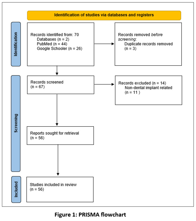 | Figure 1: PRISMA flowchart
|


